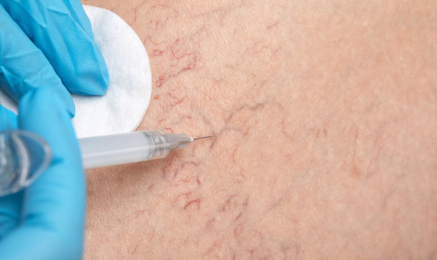
Overview
Ultrasound offers a non-invasive, radiation-free view beneath the skin. In the legs it
is used to evaluate the superficial veins, and — where appropriate — to identify the
feeder and reticular veins that supply the surface vessels patients actually notice.
What the scan shows is then used to build a personalized plan for cosmetic vein
treatment.
Dr. Ivy Lawson is double board-certified in vascular and interventional radiology.
Vascular ultrasound is not a device she added to an aesthetics menu; it is the imaging
she trained in and used in hospital practice. In Cooper City, Davie, Weston and
Pembroke Pines, that combination is unusual — most cosmetic vein treatment in the area
is offered without a physician who can perform and interpret the study.
What a vein ultrasound is used for
Evaluation of superficial veins
Identification of feeder and reticular veins when appropriate
Personalized planning for cosmetic vein treatment
Why imaging changes a vein plan
Surface spider veins are frequently the visible end of something larger. Treating the
visible vessel without understanding what supplies it is treating one part of a
pattern. Mapping the pattern first means the plan can address the sequence rather than
the symptom, and it sets a realistic expectation of how many sessions the plan is
likely to involve.
It also sorts the cosmetic from the medical. Not every leg vein finding is a cosmetic
problem. Where the scan suggests a concern that sits outside cosmetic treatment,
Dr. Lawson will say so and recommend referral or additional medical evaluation rather
than proceeding.
What happens at the appointment
The scan is non-invasive. A small amount of gel is applied and the probe is passed
over the skin — nothing is injected, and there is no radiation. Dr. Lawson performs the
study and reads it herself, and talks you through what is on the screen while you are
there. If treatment is appropriate, you leave with a plan rather than a quote.
Where it leads
Vein imaging is an assessment, not a treatment. What follows depends on what the scan
shows — most often spider vein treatment
or sclerotherapy, planned around the mapped
pattern. Patients treating the face and the legs in the same period often combine it
with facial ultrasound, and
membership pricing applies to staged treatment plans.
If you want the reasoning behind working this way,
read why we use ultrasound.
Who may benefit?
Patients seeking evaluation before spider vein treatment, patients who have had
cosmetic vein treatment elsewhere without a clear plan, and patients who would rather
know what is under the surface before committing to a course of sessions may all
benefit from ultrasound assessment when clinically appropriate.
 Ivy Wellness& Aesthetics
Book Now